Inverted EPI-LED fluorescence microscopes for GFP, FITC, TRITC and DAPI imaging — built for cell biology, tissue culture, IVF and clinical research labs. Zero warm-up, 50,000+ hour LED life, PAN India installation.

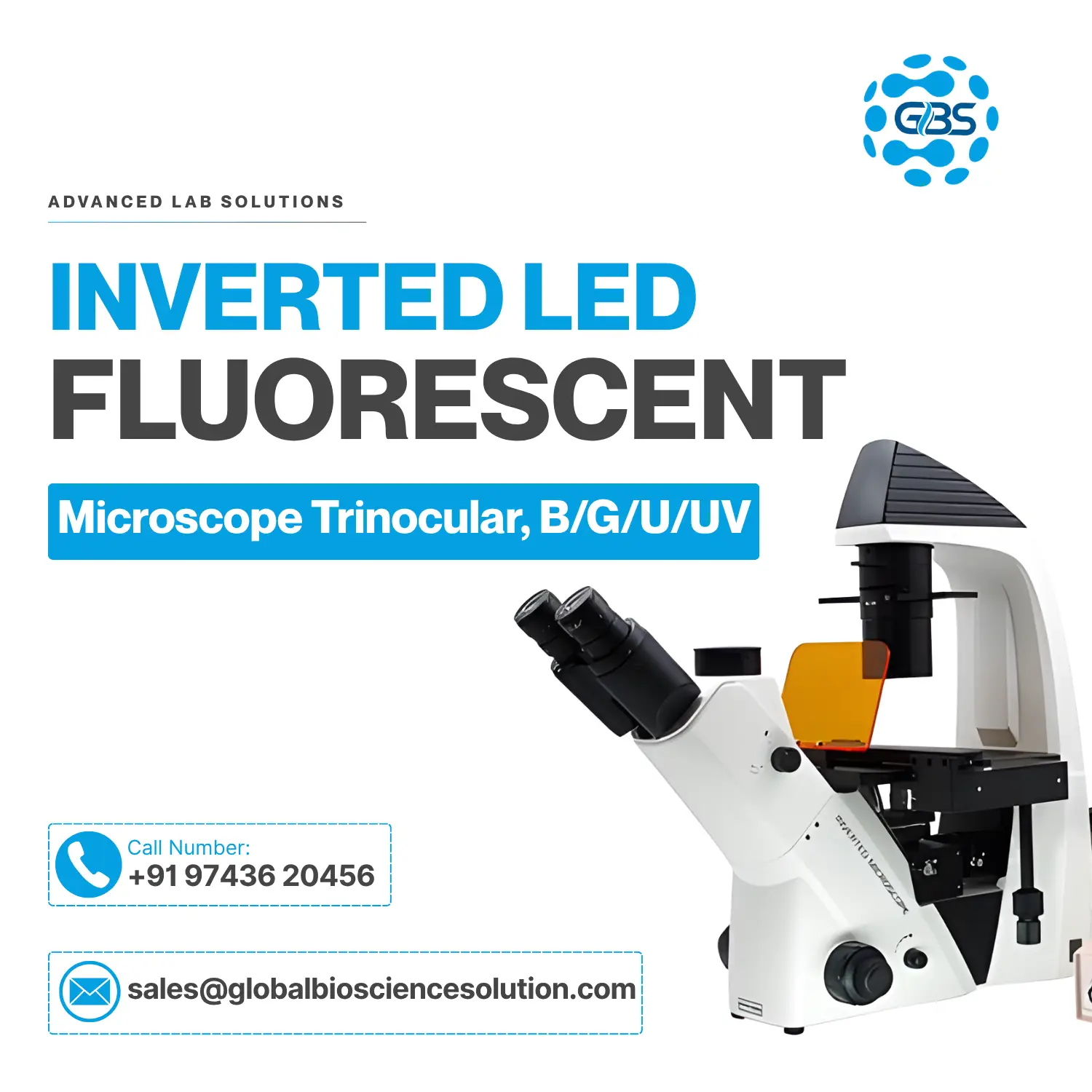
Compare filter coverage, optics and application fit across all three GBS EPI-LED fluorescence models.
| Specification | LED Fluorescent | Fluorescence (B/G) | Fluorescence (UV) |
|---|---|---|---|
| Primary Application | Tissue Culture / Cell Bio | FITC / TRITC Imaging | DAPI / UV / FITC Imaging |
| Excitation Type | EPI-LED | EPI-LED | EPI-LED |
| Filter Cubes Included | Standard | Blue, Green | Blue, Green, UV |
| Head | Trinocular | Trinocular | Trinocular |
| Optics Type | LWD Plan Achromatic | LWD Fluor Plan | LWD Fluor Plan |
| Phase Contrast | ✔ | ✔ | ✔ |
| Tier | Professional | Advanced | Advanced |
| Quote | Quote | Quote |
GBS LED fluorescence microscopes support imaging workflows across research, diagnostics and biotechnology.
Protein localization & live-cell imaging
Bacterial & fungal fluorescent labeling
Viral infection tracking studies
Neuronal marker & circuit imaging
Differentiation & marker expression
High-content screening assays
Immunofluorescence tissue analysis
IVF, embryology & screening labs
Genetic marker & expression studies
Chlorophyll & reporter gene imaging
A fluorescence microscope is an optical microscope that uses specific wavelengths of light to excite fluorescent molecules within biological samples. Unlike conventional brightfield microscopes, fluorescence microscopes detect the light emitted by fluorophores such as GFP, FITC, TRITC, DAPI and Alexa Fluor dyes, letting researchers visualize specific proteins, cells and molecular structures against a dark background.
Modern LED fluorescence microscopes provide stable illumination, a longer optical lifespan, minimal maintenance and safer operation compared with traditional mercury lamp systems — which is why LED excitation has become the standard for new lab installations.
This is one of the most searched comparisons in fluorescence microscopy — here's how the two illumination types compare.
| Factor | LED Illumination | Mercury Lamp |
|---|---|---|
| Power Consumption | Low (single-digit watts) | High (100W+ arc lamp) |
| Warm-Up Time | None — instant on | 10–30 minutes |
| Lifespan | 50,000+ hours | 200–300 hours |
| Safety | No mercury, no UV leakage | Toxic mercury, requires careful disposal |
| Maintenance | Minimal, no bulb replacement | Frequent bulb changes & alignment |
| Running Cost | Low | High (bulbs + downtime) |
| Environmental Impact | Low, no hazardous waste | Hazardous mercury waste |
Match your excitation filter to the fluorophore or stain used in your protocol.
| Fluorophore | Excitation | Emission | Filter Cube |
|---|---|---|---|
| GFP | 488 nm | 509 nm | Blue |
| FITC | 495 nm | 519 nm | Blue |
| TRITC | 557 nm | 576 nm | Green |
| Texas Red | 595 nm | 615 nm | Green |
| DAPI | 358 nm | 461 nm | UV |
| Hoechst | 350 nm | 461 nm | UV |
| Cy3 | 550 nm | 570 nm | Green |
| Cy5 | 649 nm | 670 nm | Red (optional) |
| Alexa Fluor 488 | 490 nm | 525 nm | Blue |
Match your primary application to the recommended GBS model below.
Live cell monitoring inside dishes and well plates with standard fluorescence needs.
Professional Model →Dual-channel labeling for antibody-tagged proteins using FITC and TRITC dyes.
Blue/Green Model →UV-excited nuclear counterstains alongside standard Blue/Green channels.
UV Model →| Feature | Brightfield | Fluorescence |
|---|---|---|
| Contrast | Low | High |
| Living Cells | Good | Excellent |
| Protein Imaging | ✘ | ✔ |
| GFP Detection | ✘ | ✔ |
GBS fluorescence microscopes are installed and serviced across leading Indian institutions and private labs.
Fluorescence imaging setups deployed across IIT research departments for cell biology studies.
LED fluorescence systems supporting advanced life-science research programs.
Government research labs using GBS EPI-LED microscopes for applied biology.
Diagnostic and biotech labs relying on GBS fluorescence systems for daily throughput.
Teaching and research departments across Karnataka and beyond.
Clinical and IVF labs using inverted fluorescence microscopy for embryology.
Answers to the questions researchers and lab managers ask most often about fluorescence microscopy.
Fluorescence microscopy is an imaging technique that uses specific wavelengths of light to excite fluorescent molecules (fluorophores) in a sample, then captures the longer-wavelength light they emit, revealing specific proteins, cells or structures against a dark background.
Light from an LED source passes through an excitation filter, reflects off a dichroic mirror onto the sample, and excites fluorophores. The emitted light returns through the mirror and an emission filter to reach the eyepiece or camera, isolating only the fluorescent signal.
GFP (Green Fluorescent Protein) is a naturally fluorescent protein used as a genetic marker. It excites near 488 nm and emits green light near 509 nm, visible under a Blue-filter fluorescence microscope.
FITC (Fluorescein Isothiocyanate) is a synthetic green-emitting dye used to label antibodies and proteins. It excites around 495 nm and emits around 519 nm under a Blue filter cube.
TRITC (Tetramethylrhodamine) is an orange-red fluorescent dye often paired with FITC for dual-labeling. It excites around 557 nm and emits around 576 nm under a Green filter.
DAPI is a blue-fluorescent dye that binds strongly to DNA and is used to stain and visualize cell nuclei. It requires UV excitation (around 358 nm) and emits near 461 nm.
UV fluorescence microscopy uses a UV excitation filter cube (roughly 330–380 nm) to excite dyes such as DAPI and Hoechst, commonly used for nuclear staining.
Yes. When bacteria are labeled with fluorescent stains or express fluorescent proteins, a fluorescence microscope can detect and localize them with high contrast against unlabeled background material.
Fluor-corrected, long working distance (LWD) plan objectives are recommended, as they preserve signal brightness and allow imaging through culture vessel plastic.
Numerical aperture describes an objective's light-gathering ability and resolving power. Higher NA objectives capture more emitted light, which is especially valuable in low-signal fluorescence work.
LED illumination is generally preferred over mercury lamps because it needs no warm-up time, lasts over 50,000 hours, uses less power, runs cooler and avoids hazardous mercury disposal.
Entry-level inverted LED fluorescence microscopes in India typically start in the low lakhs and scale up with additional filter cubes, camera integration and optics quality. Contact GBS Microscope for a configuration-specific quotation.
IVF and embryology labs typically choose an inverted LED fluorescence microscope with a long working distance condenser and Blue/Green filters, allowing direct imaging of live cultures inside dishes without disturbing the sample.
The GBS Inverted LED Fluorescent Microscope (Professional tier) is purpose-built for tissue culture, with an LWD condenser for imaging directly inside flasks and well plates.
For most Indian research labs, an inverted EPI-LED model with Blue, Green and UV filter cubes offers the broadest fluorophore coverage across FITC, TRITC and DAPI protocols.
Minimize exposure time, reduce excitation intensity where possible, use fresh fluorophores, and limit the sample's total light exposure by focusing under brightfield before switching to fluorescence.
Start with a longer exposure time and moderate gain rather than high gain alone, since fluorescence signals are typically dim; adjust based on the specific fluorophore's brightness and photostability.
Yes. GBS Microscope provides PAN India installation, calibration, AMC, training and application support for every fluorescence microscope sold.
Yes. LED excitation runs cool and avoids the intense heat and UV leakage associated with mercury lamps, making it safer for extended live-cell imaging sessions.
A standard Blue excitation filter cube covers both GFP (488/509 nm) and FITC (495/519 nm), since their excitation and emission wavelengths are closely matched.
Our imaging specialists respond within 24 hours · PAN India delivery & installation included