High-performance inverted microscopes for live-cell imaging, tissue culture, IVF, metallurgy and advanced fluorescence. Observe specimens directly in culture flasks and petri dishes. PAN India delivery & installation.


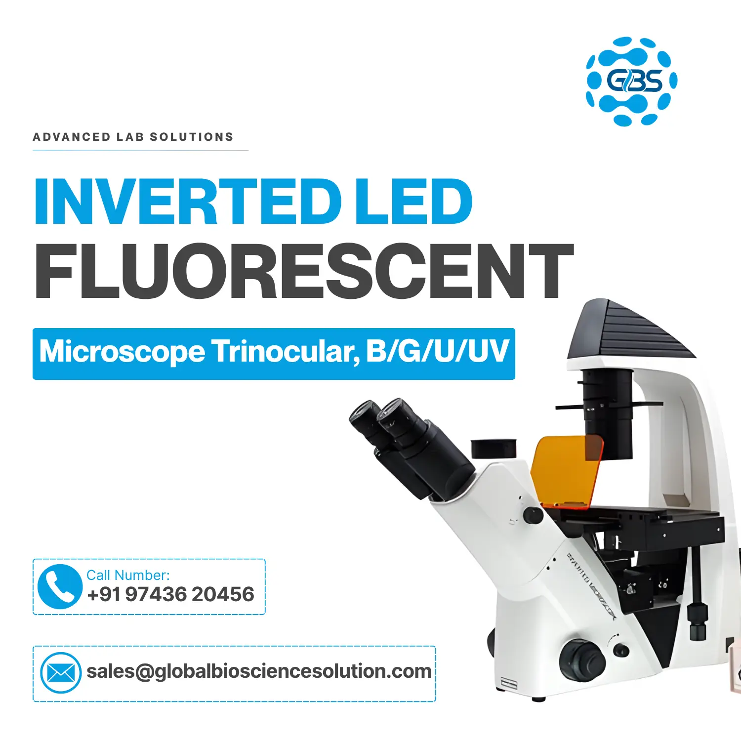
Compare optics, illumination and filter coverage across all five GBS inverted microscope models.
| Specification | Bio Transmit/Reflect | Metallurgical Reflected | Fluorescence (B/G) | Fluorescence (UV) |
|---|---|---|---|---|
| Application | Live Cell / Tissue Culture | Metal & Wafer Inspection | FITC/TRITC Imaging | DAPI/UV Imaging |
| Optics Type | LWD Plan Achromatic | Infinity LWD M-Plan | LWD Fluor Plan | LWD Fluor Plan |
| Head | Trinocular | Trinocular | Trinocular | Trinocular |
| Phase Contrast | ✔ | — | ✔ | ✔ |
| Fluorescence Filters | — | — | Blue & Green | Blue, Green & UV |
| Polarizing Kit | — | ✔ | — | — |
| Illumination | Transmit & Reflected | EPI Reflected Halogen/LED | EPI-LED Fluorescence | EPI-LED Fluorescence |
| Tier | Professional | Advanced | Advanced | Advanced |
| Quote | Quote | Quote | Quote |
An inverted microscope is a microscope built with its optical layout flipped relative to a standard laboratory microscope: the objective lenses sit below the stage, while the illumination source and condenser sit above it. This arrangement lets researchers view a sample from underneath, through the base of an open container such as a petri dish, culture flask or multi-well plate.
Why is it called "inverted"? The name simply describes the reversed position of the objective and light source compared with a conventional upright microscope, where the objective looks down onto a slide from above.
When should it be used? An inverted microscope is the right choice whenever a specimen is living, submerged in liquid medium, or held in a vessel too deep or too large to place under a standard upright stage — cell cultures, IVF embryos, and even metal or semiconductor samples resting flat on a bench all fall into this category.
How is it different from an upright microscope? An upright microscope images thin, fixed specimens on glass slides from above; an inverted microscope images live or bulky specimens from below without needing to mount them on a slide at all.
Which industries use it? Cell biology and IVF labs, pharmaceutical and biotech research, university life-science departments, and — thanks to reflected-light metallurgical variants — metal, semiconductor and materials-science inspection labs.
The objective's position below the stage is what makes live-cell and culture-vessel imaging possible.
Because the objective is positioned beneath the stage, it can focus upward through the clear plastic or glass base of a dish without interference from the liquid medium above. This means a live-cell culture can be observed, measured and imaged for hours or days at a time, without ever removing it from its incubation vessel — something a standard upright microscope cannot do. Long working distance (LWD) objectives are used specifically to keep the lens far enough from the specimen to accommodate the thickness of the dish and the depth of the medium.
One of the most searched comparisons in microscopy — here's how the two layouts differ in practice.
| Feature | Inverted | Upright |
|---|---|---|
| Objective Position | Bottom | Top |
| Best For | Live cells | Fixed slides |
| Cell Culture | ✔ | ✘ |
| Petri Dishes | ✔ | ✘ |
| Tissue Sections | Limited | Excellent |
| Metallurgy | ✔ | Limited |
Live monitoring in flasks & dishes
Embryo observation & grading
Tumor cell growth studies
Differentiation monitoring
Compound screening assays
Neuronal culture imaging
Immune cell interaction studies
Live microorganism observation
Flat metal sample analysis
Wafer & chip surface checks
Microstructure analysis
Fracture & defect inspection
| Objective | Working Distance | Best Use |
|---|---|---|
| 4X | Long | Large samples, overview scanning |
| 10X | Medium | Cell culture, general screening |
| 20X | Medium | Live cell detail imaging |
| 40X | Short | Fine cellular detail |
| 60X | Oil | High-resolution research imaging |
| Light Source | Lifespan | Heat | Maintenance | Cost | Ideal For |
|---|---|---|---|---|---|
| LED | 50,000+ hrs | Low | Minimal | Low | Brightfield, phase, general use |
| Halogen | 1,000–2,000 hrs | Moderate | Periodic bulb change | Low | Standard transmitted light |
| Mercury Lamp | 200–300 hrs | High | Frequent bulb & alignment | High | Legacy fluorescence setups |
| Fluorescence LED | 50,000+ hrs | Low | Minimal | Moderate | GFP, FITC, TRITC, DAPI imaging |
Answer these questions in order to land on the right GBS model for your lab.
Routine live-cell monitoring in flasks, dishes and well plates.
Biological Model →FITC, TRITC, GFP or DAPI imaging inside live culture vessels.
Fluorescence Model →Opaque, flat samples such as metals, alloys and semiconductor wafers.
Metallurgical Model →How GBS inverted microscopes are applied across research, clinical and industrial settings.
Daily live-cell health checks in academic and biotech labs without disturbing incubation.
Direct imaging of embryos inside culture dishes for grading and selection.
Reflected-light inspection of wafer surfaces for defects and contamination.
Quality control imaging of cell-based assays during drug development.
Metallurgical inspection of fractured or failed components for root-cause analysis.
Long-term tumor cell growth and treatment-response imaging.
Answers to the questions researchers and lab managers ask most often about inverted microscopes.
An inverted microscope has its objective lenses below the stage and its light source above, letting you view samples in open containers such as petri dishes and flasks directly from underneath.
An inverted microscope lets you observe live cells and cultures without removing them from their incubation vessel, and it handles bulky or opaque samples like metal blocks that can't sit on a slide.
An inverted microscope images from below through open dishes and is suited to live cells; an upright microscope images from above on fixed glass slides and is better suited to thin tissue sections.
Yes — this is their primary strength. The objective focuses through the base of the culture vessel, so cells can be observed continuously without disturbing the liquid medium.
LWD refers to objectives and condensers designed with extra clearance from the lens to the sample, needed to focus through the thickness of a culture dish and its liquid medium.
Phase contrast is an optical technique that converts subtle differences in light phase, caused by transparent structures like cells, into visible contrast differences without staining the sample.
An inverted biological microscope with LWD optics and phase contrast is the standard choice for IVF labs, allowing direct embryo observation inside culture dishes.
The GBS Inverted Microscope with Transmit & Reflect Light (Professional tier) is purpose-built for tissue culture, combining LWD optics with phase contrast.
Yes. Inverted fluorescence models add EPI-LED excitation and filter cubes to the standard inverted optical path, enabling GFP, FITC, TRITC and DAPI imaging directly in live cultures.
Standard LWD plan achromatic objectives (4X–40X) cover most biological work, while LWD Fluor-corrected objectives are used for fluorescence, and Infinity M-Plan objectives are used for metallurgical inspection.
Working distance is the gap between the front of the objective lens and the sample when it's in focus. Longer working distances are needed to image through the base of taller culture vessels.
A basic biological inverted microscope typically starts in the low lakhs; fluorescence and metallurgical variants cost more depending on optics, filters and camera integration. Contact GBS Microscope for a specific quotation.
The right model depends on your application — GBS offers biological, fluorescence and metallurgical inverted models, each optimized for a different type of sample and workflow.
Yes. Inverted metallurgical microscopes use reflected light instead of transmitted light, allowing flat, opaque samples like metals and semiconductor wafers to simply rest on the stage for inspection.
Standard culture dishes and flasks with clear plastic or glass bases work well; very thick or textured plasticware can reduce optical clarity at higher magnifications.
Yes. GBS Microscope provides PAN India installation, calibration, AMC, training and application support for every inverted microscope sold.
Yes. All GBS inverted models feature a trinocular head with a dedicated camera port compatible with HDMI or USB digital cameras and imaging software.
Biological models use transmitted light through a translucent sample from below; metallurgical models use reflected light bounced off an opaque sample's surface, since light cannot pass through metal.
Yes, an inverted metallurgical microscope with reflected light and a flat, oversized stage is well suited to inspecting wafer surfaces for defects and contamination.
Looking for dedicated fluorescence imaging? See our full Fluorescence Microscopes range.
Our experts respond within 24 hours · PAN India delivery & installation included